PLANMECA
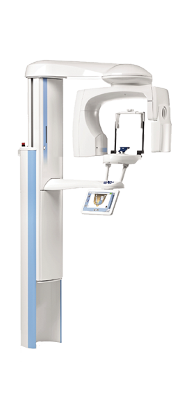
We are pleased to announce that Dr. Kauffman is the first in the state of Georgia to add the ProMax X-ray machine to our office. This machines technology brings new possibilities for treatment planning and maximizes the safety of our imaging procedures. This unit complies with the best practices in dentistry by following the American Dental Association's ALARA (As Low As Reasonably Achievable) radiation principle to minimize the effective radiation dose to our patients. The unique features of this unit allow us to choose what area of the teeth and facial anatomy we need to capture, which significantly reduces any unnecessary exposure. Our staff can also adjust the unit's settings, allowing us to capture only the details we need. We will be using this unit with state-of-the-art software applications for the best possible care using
Ultra Low Dose Imaging and Diagnostics
Dr. Kauffman is a stalwart advocate of the use of 3D x-ray imaging in dentistry. This large FOV (field of view) enables Dr. Kauffman to evaluate airways, orthodontic cases, TMJ issues, endodontic therapy, and implant planning.
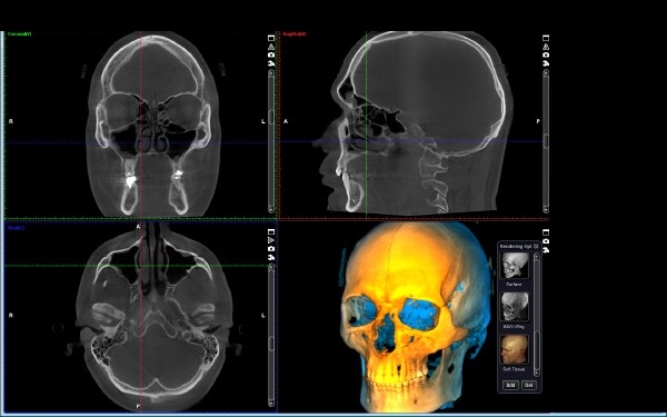
3D x-ray imaging is known to the medical world as Cone Beam Volumetric Tomography (CBVT). It has been adapted to the dental field by reducing volume size and drastically reducing radiative exposures to produce the images. In Dr. Kauffman’s practice, CBVT has been used to help plan implant surgeries to ensure ideal placement is achieved.
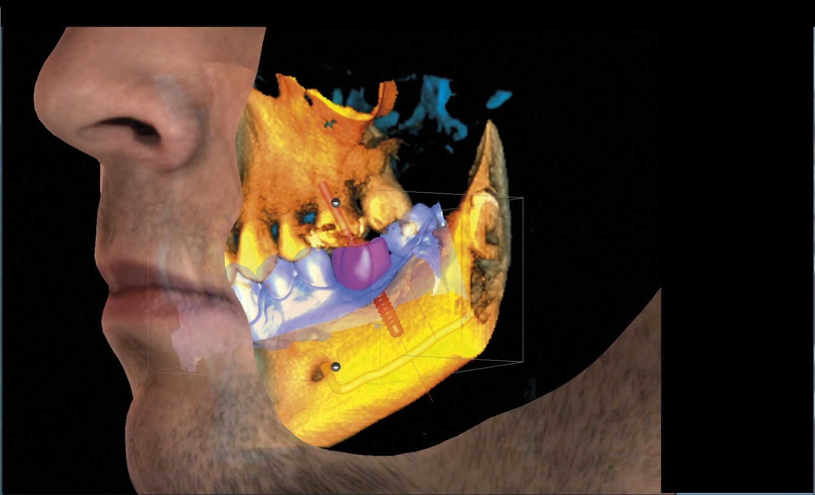
The CEREC CAD/CAM technology enables Dr. Kauffman to take 3-dimensional models of patients’ teeth and relevant oral anatomy. These 3D models can be integrated into a 3D CBVT image to enable Dr. Kauffman, to map nerves and digitally place an implant:
In addition to implant planning, CBVT offers many other advantages over traditional imaging, but especially in the area of oral pathology detection. Dr. Kauffman uses his 40+ years of dental experience and occasionally makes use of a 3 rd party radiologist to assist in certain diagnosis.
Obstructive Sleep Apnea (OSA) is a potentially harmful disorder caused by and airway that is naturally too small. Traditional forms of dental imaging would not be able to capture an image of a patient’s airway. With CBVT imaging we are able to not only capture an image of the airway, but also perform a preliminary evaluation using special imaging software:
3D CBVT technology represents part of the future of dentistry. It is improving pathology detection, implant and restorative case planning, and the effectiveness of twice yearly exams. If you are ready for a comprehensive dental exam, in a practice that is on the cutting edge of dental technology, then