(MORE PICS BELOW ARTICLE)
POSTERIOR ARTHROSCOPY
(Ankle and Subtalar Joint)
By Robert H. Sheinberg, D.P.M., D.A.B.F.A.S., F.A.C.F.A.S.
What is it?
A minimally invasive procedure that is done by going into the back of the ankle and the subtalar joint with very small incisions to clean out any joint, ligament, cartilage or tendon damage.
Why?
Traditional surgery in the past included making small or long incisions to access joints and tendons. Arthroscopy can be done with two small incisions 1/6” in length, giving greater access to areas very difficult to visualize. More can actually be done through very small incisions with an arthroscope because visualization is greatly enhanced.
Benefits:
- Greater access to all the injured areas with very small incisions.
- Minimally invasive.
- Less risk of complications.
- Less risk of scarring.
- Less risk for infection.
- Ability to immediately move the foot and ankle the next day after surgery to help restore range of motion, strength and stability.
- Able to resume sporting activities including dance, football and basketball over a 1-2 week period of time in most cases.
Conditions Treated:
- Fractures in the back of the ankle and foot.
- Impingement syndrome, especially in dancers soccer and basketball players.
- Synovitis, which is an inflammation of the joint lining.
- Scarring in the back or side of the ankle or foot.
- Tenosynovitis of the flexor hallucis longus tendon.
- Treatment for “OS TRIGONUM SYNDROME”.
- Treatment for enlarged lateral talar tubercle.
Prognosis:
- Excellent in almost all cases if the damage is limited to the soft tissue or fragments of bone or cartilage.
- Better but a guarded prognosis if there is extensive osteoarthritis to the foot or ankle.
********************************************************************
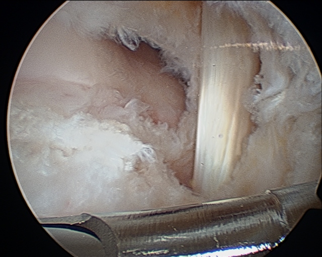
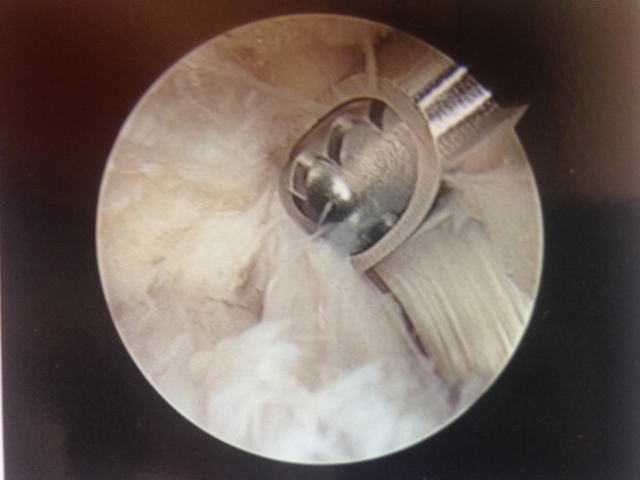
Posterior arthroscopy
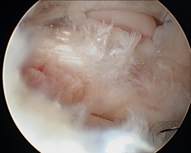
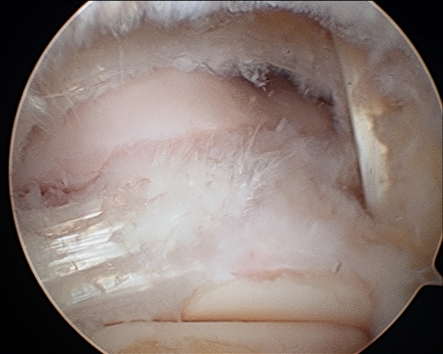
Scar tissue in the back of the ankle
After debridement, the FHL can be on the right-hand side
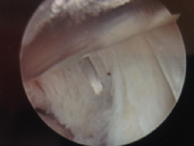
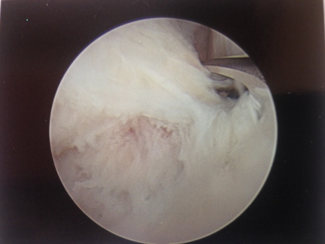
The ankle joint can be seen at the top of the screen clear of any scar tissue or debris
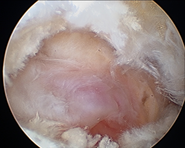
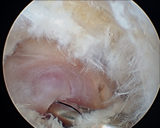
Posterior scope with removal ganglion cysts
The ganglion is the mass with the reddish hue inside the back of the joint
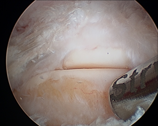
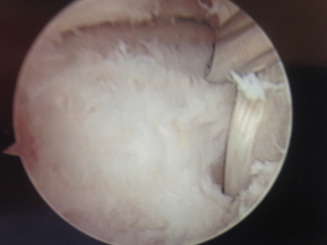
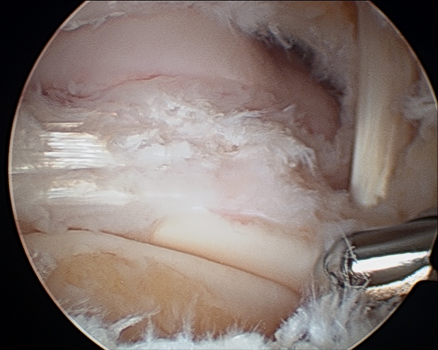
The ankle and subtalar joint visualized post debridement
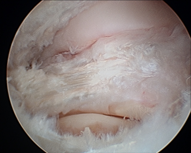
A portion of the posterior ankle ligament complex
The cleaned out ankle joint
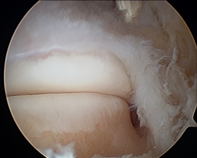
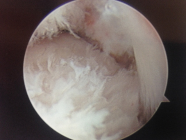
The cleaned out ankle and subtalar joint easily visualized. The FHL tendon is noted at the top right of the picture
The FHL tendon after debridement