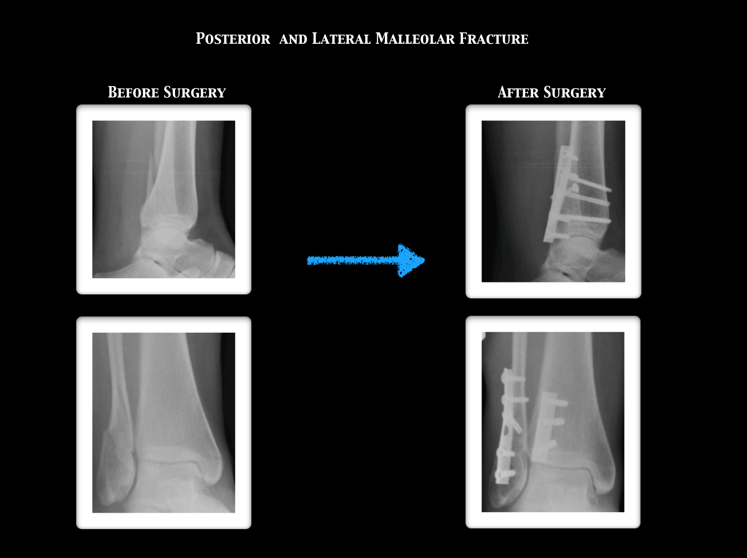
Posterior Malleolar Fractures
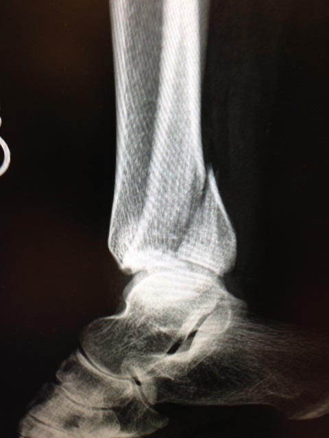
CT scan and Xray sagittal views of severly displaced posterior tibial malleolar fracture with comminution. This type of injury can cause significant problems in the future with disability if not treated with surgery to reduce the fracture and fixate.
CT scan sagittal views of minimal displaced posterior tibial malleolar fracture.

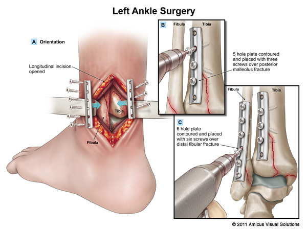
Trimalleolar fractures involve fracture of the medial and lateral malleoli along with a fracture of the posterior lip of the tibial plafond; (posterior malleolar fracture). Fracture results from avulsion by the posterior tibiofibular ligament at its site of attachment to the tibia. The irregularity in the tibial articular surface of the tibia is brought against the weightbearing surface of the talus and with motion and weightbearing severe DJD develops.
X-ray of Non Displaced Posterior Malleolar Fracture
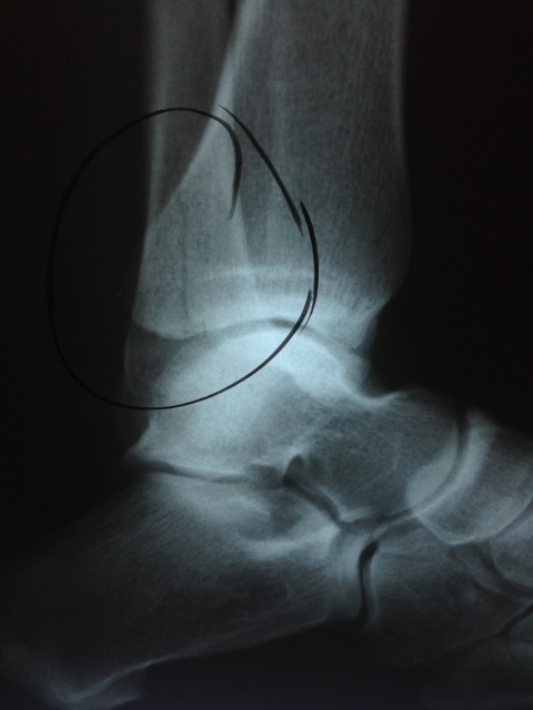
X-ray of Displaced Posterior Malleolar Fracture
Indications for surgery:
- 25% of the posterior articular surface is involved
- Fragment must have a tibial articular surface that is large enough to provide a stable weightbearing surface
- Fracture is displaced more than 2 mm
- Ankle fracture dislocation with posterior malleolar fracture
- Posterior subluxation of the talus
- Even the slightest posterior subluxation of the talus on the articular surface of the tibia is not acceptable
- If fracture prevents reduction of the fibula
- Valgus tilt of the talus in post-reduction film may be a relative indication
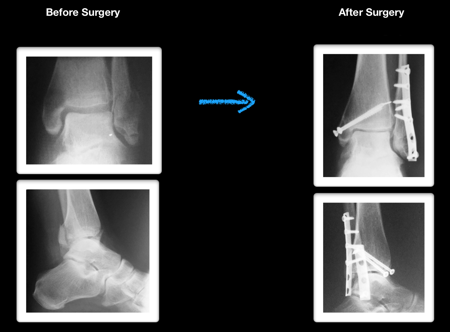
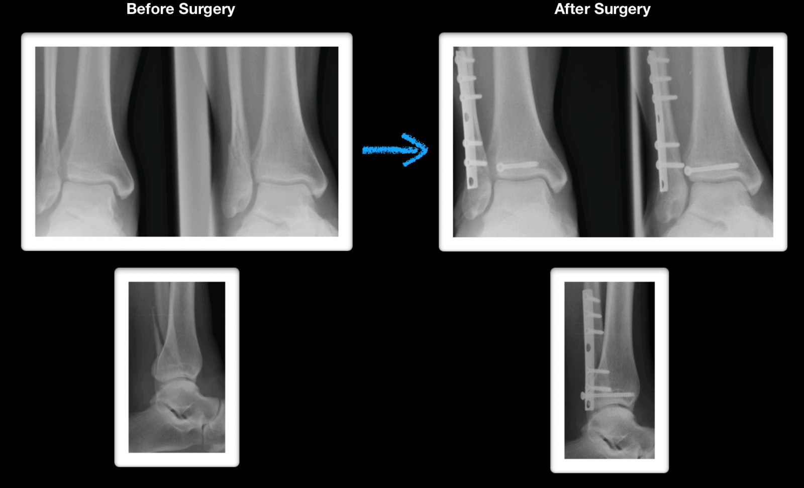
Pre and Post Operative X-Rays
Xray and CT scan of displaced medial and posterior malleolar fracture with ORIF
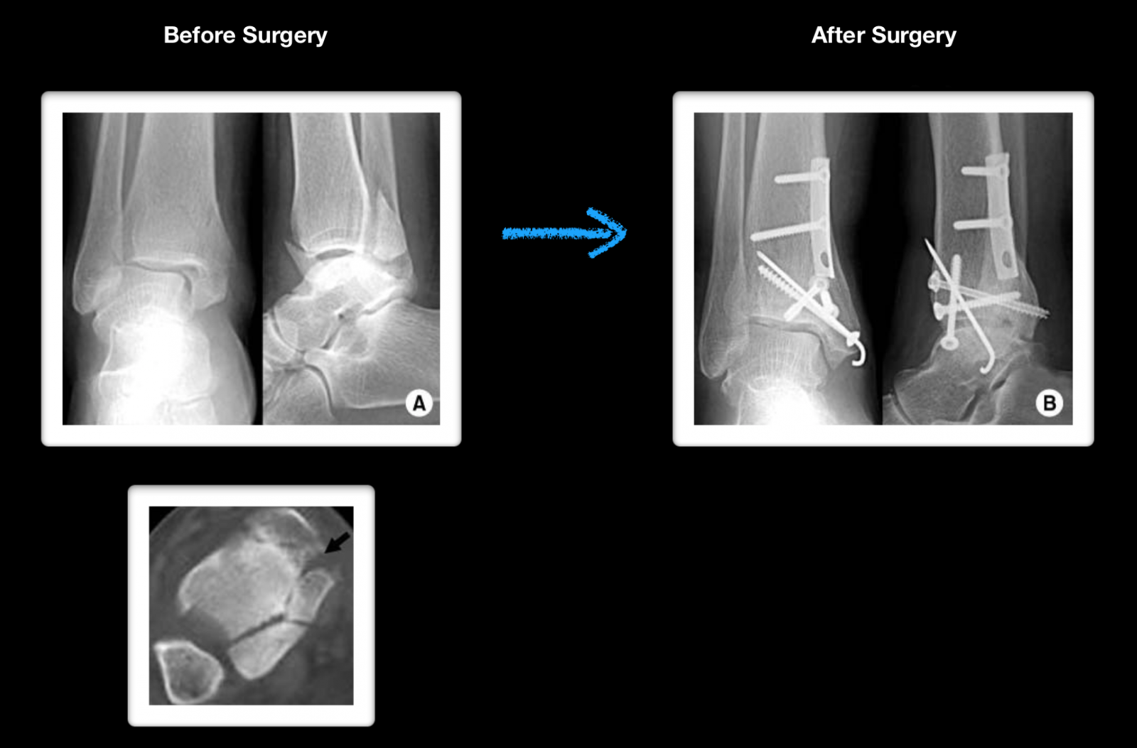
Preop and Postop ORIF Posterior Malleolus Fracture with associated Fibular fracture
Preop and Postop Trimalleolar Fracture with Fixation of Posterior Malleolus Fracture
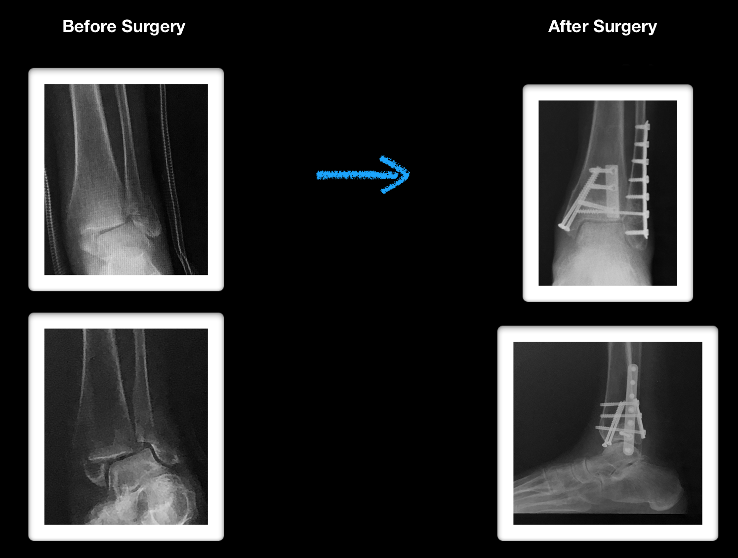
Pre and postop Trimalleolar Fracture without FIxation of Posterior Malleolus