SESAMOID FRACTURES
Robert H. Sheinberg, D.P.M., D.A.B.F.A.S., F.A.C.F.A.S.
Sesamoids:
The sesamoids are two small bones in the ball of the foot that go underneath
the first metatarsal. These bones are almost completely within the tendons
that control the function of the big toe joint. When somebody walks, the
sesamoids assist the small muscles in the ball of the foot to firmly plant
the big toe on the ground. This allows us to effectively weightbear on the
big toe and push off during sports and activities. Injuries to the sesamoid
can be very painful and many athletes limit their running ability and even
their athletic career. Sesamoid injuries are also very common in dancers
because of the excess stress that is placed on the ball of the foot. They
are also common in running athletes, especially a skilled position in
football players because of their excessive running on the ball of the
foot.
Stress Fractures:
Stress fractures are relatively common in the sesamoid in athletes or
dancers that are required to be on the ball of the foot. X-rays are often
negative and MRIs are needed to evaluate the extent of the injury. Treatment
is often nonweightbearing in a boot to allow the bone to heal. It can take
many months before the bone heals completely and utilization of an orthotic
following the healing would further unload the sesamoid.
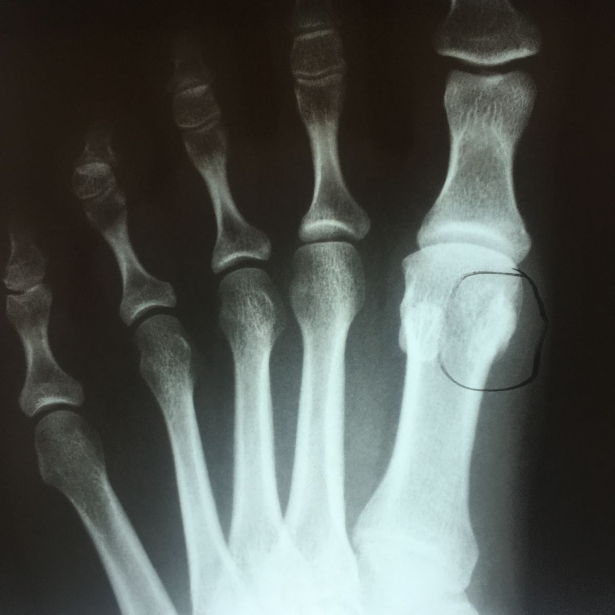
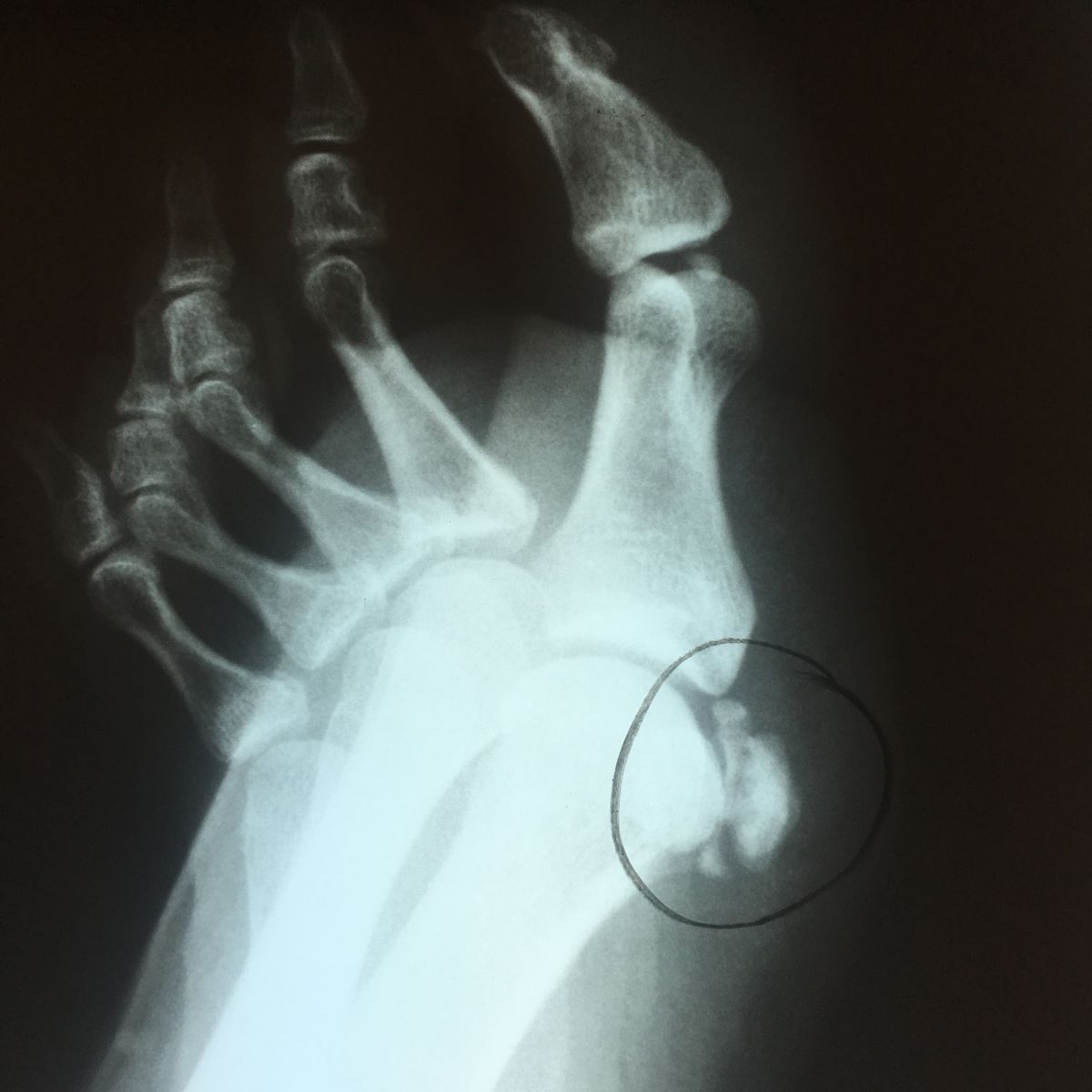
Comminuted Fractures:
Fractures to these bones can occur from a single traumatic event. This would
be common in somebody falling from a height and landing on the ball of the
foot. Fractures of the sesamoids can be nondisplaced or moderately
displaced. Displaced fractures to this bone are often difficult to treat.
Surgery is often necessary to remove one or more of the fracture fragments
and allow the person or athlete to return to sporting activities.
Nondisplaced fractures can be treated conservatively. This requires a boot
nonweightbearing for a period of 6-8 weeks. Healing of these fractures can
be very prolonged. Following immobilization, an orthotic put in the person’s
shoe would assist the patient in unloading the area to maximize the healing
process.
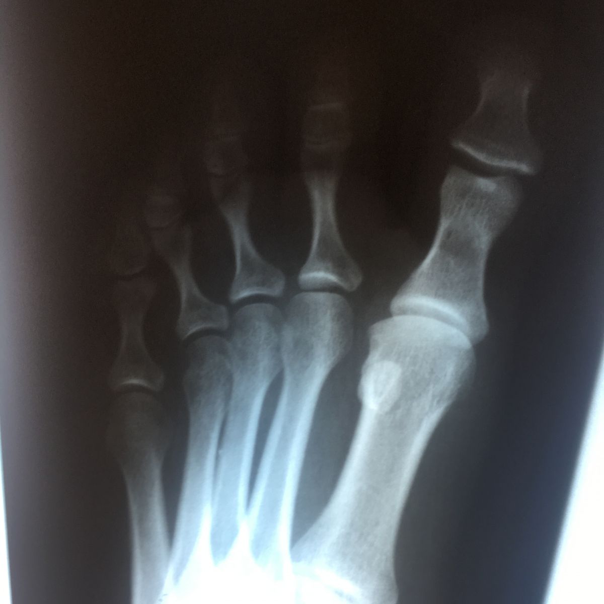
Synchondrosis Fractures:
The sesamoids can be normally in two or more pieces. These pieces are
connected by cartilage. This is referred to as a synchondrosis. Cartilage
has a very poor blood supply to the area. When an injury occurs to the
synchondrosis it may appear as a fracture on an x-ray but it is not a
fracture of the bone but a fracture of the cartilage connecting the bones.
Injuries to the synchondrosis can be very difficult to treat. X-rays may be
taken on the unaffected side to compare. However, both sides may have what
appears to be sesamoids in multiple pieces. When pain persists, then an MRI
may be necessary to fully evaluate the injury. We often see bone marrow
swelling on two or more bone segments, which indicates the injury to that
area. Clinically, there is pinpoint tenderness to the bone on the ball of
the foot. Swelling may or may not be present. There will be difficulty
walking in long strides or running on the ball of the foot. Dancing can
become near impossible because of the pain that patients may experience.
Treatment includes immobilization in a boot and often nonweightbearing to
allow the area to heal. These injuries will most often heal with
conservative care but it may take many months before it goes on to complete
healing. During the healing process, x-rays will not change, as the bones
will not grow back together because of the cartilage that is between them.
We can evaluate the healing based on the patient’s pain that they are
experiencing while walking and examining them to check for tenderness. Once
tenderness and pain are diminished, then an orthotic is made to go in the
patient’s shoes or sneakers to unload the metatarsal further and allow the
patient to return to sports and activities as desired. In some cases, these
injuries do not heal and one or more of the pieces of bone on the ball of
the foot needs to be removed. A small incision is placed on the inside of
the first metatarsal joint and a piece of the bone is removed. The soft
tissue is repaired and immobilization for 3-6 weeks is necessary to allow
the area to heal completely. The long-term prognosis is usually excellent.
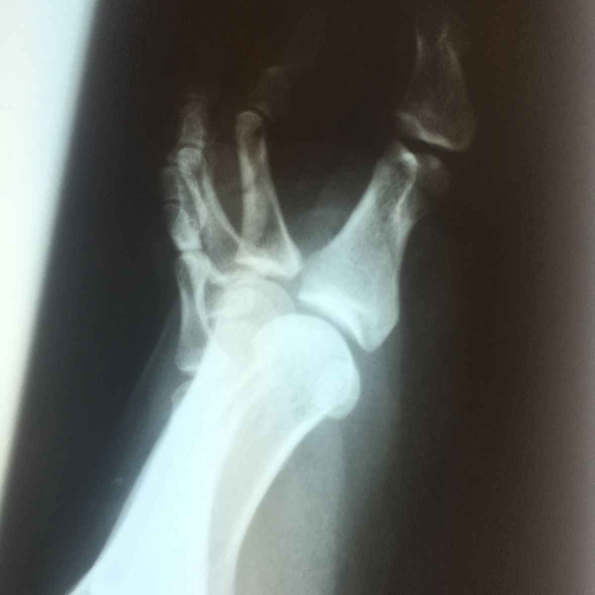
Avascular Necrosis:
Avascular necrosis can develop in one or both of the sesamoids. This occurs
when the blood supply to the bone has been disrupted and the bone fails to
revascularize. The bone essentially dies and causes chronic pain to the ball
of the foot. The sesamoid articulates with the first metatarsal bone. As the
big toe moves up and down the sesamoid runs forward and backward. Arthritis
can develop at the articulation between the two bones, causing pain with
walking and certainly with running. X-rays may be of some benefit to
evaluate the sesamoid injury. However, MRIs are almost always mandatory to
evaluate the full extent of the injury. If the bone has died and pain
persists causing an inability to weightbear for three or more months,
surgery may be necessary to remove the sesamoid and restore the person’s
ability to go back to sports and activities.
Arthritis:
Arthritis of the sesamoid-metatarsal articulation is relatively common.
Arthritis is a degenerative process where the cartilage of the bone starts
to wear down. When the cartilage wears down between the sesamoid and the
metatarsal, bones start to rub against each other and chronic pain and
swelling often develop. This can occur from one traumatic event such as a
fall from a height. It can also occur from low-grade stress to the ball of
the foot that persists over time. It is very common to see arthritis in the
area in patients with bunions or arthritis of the big toe joint. At times
the arthritis is relatively asymptomatic and in some cases it can become
very painful, limiting all activity. MRIs and occasionally CT scans are
necessary to fully evaluate the injury. If pain persists despite
conservative care (anti-inflammatories, injections, and orthotics), then
surgery would be necessary to remove the sesamoid.
Pre and Postop Tibial Sesamoidectomy