Dental 3D imaging is commonly used for diagnosing and treatment planning of cases that involve:
- surgical planning for impacted teeth.
- diagnosing temporomandibular Joint Disorder (TMD).
- accurate placement of dental implants.
- evaluation of the jaw, sinuses, nerve canals and nasal cavity.
- detecting, measuring and treating jaw tumors.
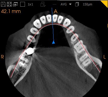
- determining bone structure and tooth orientation.
- locating the origin of pain or pathology.
- hard to locate tooth fractures.
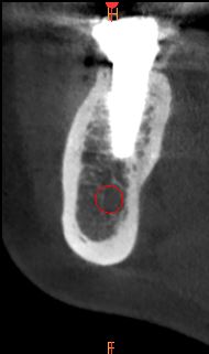
- hard to locate canal systems.
Doctor Benefits
- One scan provides you with significantly more information about your patient's anatomy and bone structure than traditional 2D films.
- These distortion free images display implant site assessment, TMJ disorders, impacted teeth, critical bone and tooth relationships, mandibular canal and difficult to see pathologies.
- Images can be easily viewed on a computer screen.
- Patient's understanding of anatomy and the treatment plan leads to better patient acceptance.
Patient Benefits
- The radiation exposure is less than a full mouth series and 95% less than a conventional CT scan.
- Much more economical than hospital based conventional CT imaging.
- It is quick, comfortable, and painless for the patient. Taken in an open upright chair, most scans are only 10 seconds.
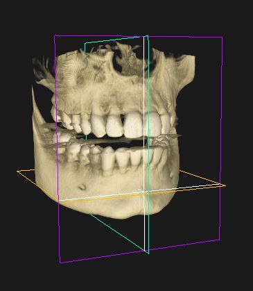
- The patient can more easily understand the problem and solution by viewing the 3D skull volume on the computer monitor.
- See how it works!