
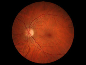
 Conventional Photo
 Optos Image
THOSE WHO HATE TO HAVE THEIR EYES DILATED
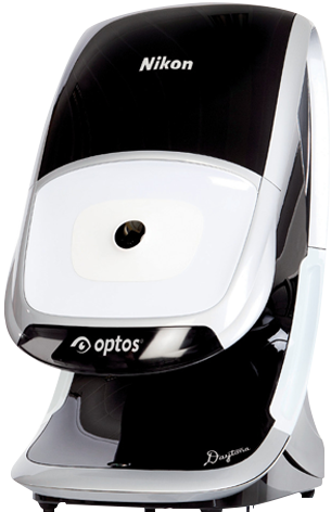
Because we are completely dedicated in your well being and the health of your eyes, we have invested in the newest cutting edge technology in retinal imaging that reduces the need to dilate your eyes. Optos is an ultra-widefield imaging system that allows us to see a 220 degree view of the back of your eye (the retina) vs the 45 degree view of prior technology.
The retina is the only place in the body that blood vessels are directly visible, therefore it can be the FIRST place to detect the following pathologies:
- Diabetes
- Hypertension
- High cholesterol
- Stroke
- Heart Disease
- Aneurysms
- Brain tumors
- Toxemia of Pregnancy
- Medication toxicity
- Autoimmune diseases
- Glaucoma
- Macular degeneration
It is very benefical with the diagnosis and evaluation of retinal tears, and detachments. Optomap is a great way to monitor your eye health, so we recommend that everyone get at least a baseline scan. We may recommend having the test done more frequently depending on your individual health status. For instance, diabetics should have this test done annually. The test is completely painless and only takes seconds to perform. It is perfect for patients of all ages, even children, so the whole family can benefit from this new technology and can feel confident they are getting the most advanced and up-to-date care.
As Optomap reduces the need for dilation, it does not totally eliminate the need to dilate. There still will be specific presentations where dilation is needed. If symptoms or test results reveal any suspicious findings, we will the need to dilate your eyes for further investigation. Talk to one of the doctors about this new and exciting technology at your next visit!
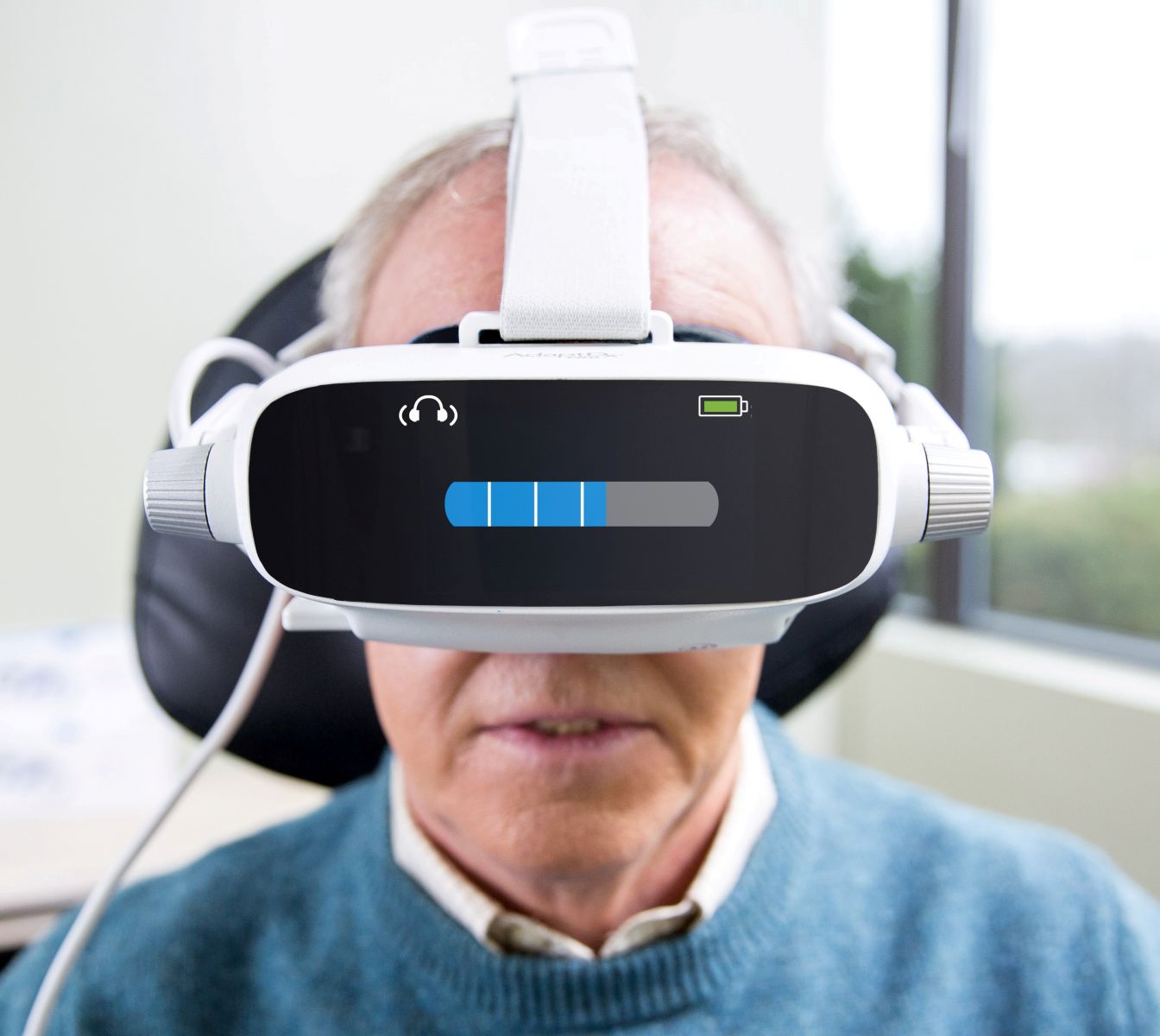
 from Maculogix
https://www.youtube.com/watch?v=RH3zugWRpAQ

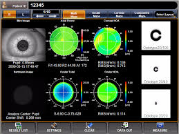
 NIDEK WAVEFRONT CORNEAL TOPOGRAPHER

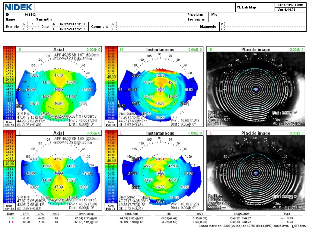
By taking over 1000 readings on the cornea, Wavefront topography gives us the advantage of designing the most comfortable, safest contact lens possible. And most importantly, it provides unparalleled accuracy when predicting LASIK outcomes. Note the vertical yellow "bow tie" on the topography demonstrating two separate curves, revealing an astigmatism. This instrument along with the KRW1 below, work in tandum to provide an invaluable amount of data, enabling us to recommend solutions that will give the clearest vision possible.
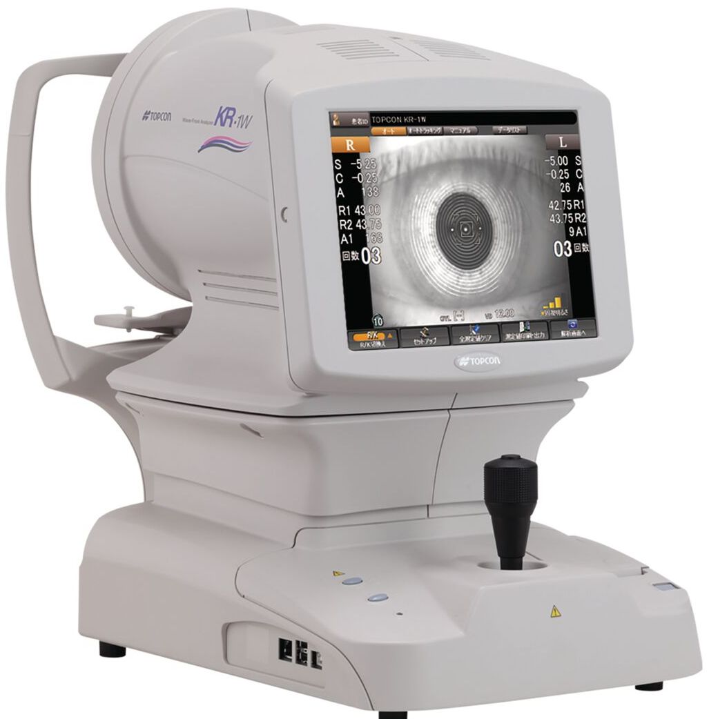
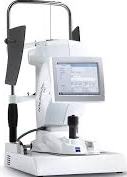
KR1-W AUTOREFRACTOR
This latest diagnostic tool combines 5 functions to fully evaluate the optical system of the eye.
- Autorefractor: Establishes the prescription required to provide the clearest vision in your spectacles
- Keratometry: Measures the curvature of the cornea for any astigmatism
- Topography: Gives the precise shape of the eye which enhances LASIK and Myopia Management results
- Aberrometry: Uncovers unwanted distortions that can not be corrected with glasses or LASIK
- Pupillometry: Measures pupil size in light and dark environments which is a cause of night halos
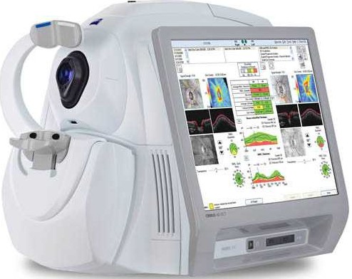
 CIRRUS 6000 OCT (OPTICAL COHERENCE TOMOGRAPHY)
This breakthrough technolgy takes a 100,000 scans per second of the structures below what is visible to the doctors eye. It can detect the earliest signs of macula degeneration and glaucoma, and diabetes years before presenting themselves in a routine examination, enabling prompt treatment before irreversible vision loss.
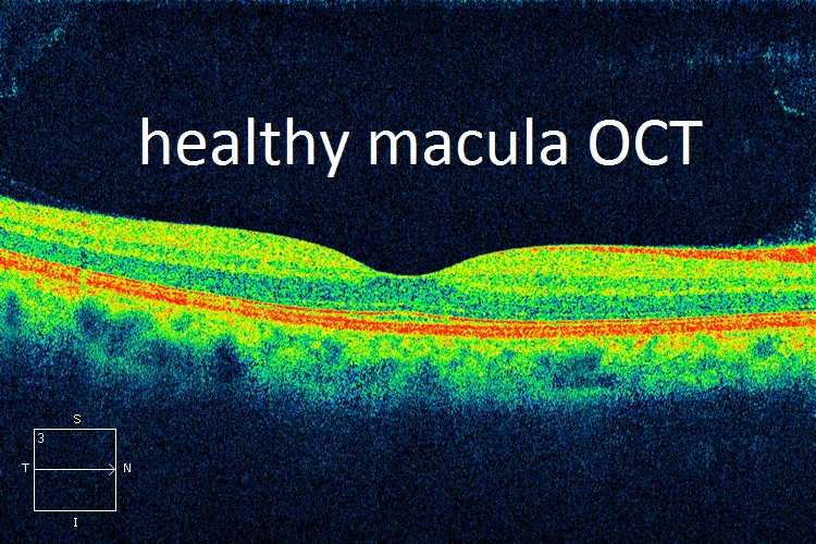
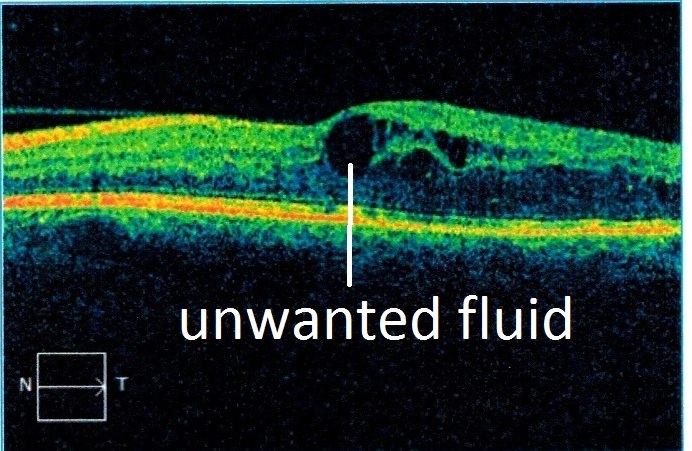
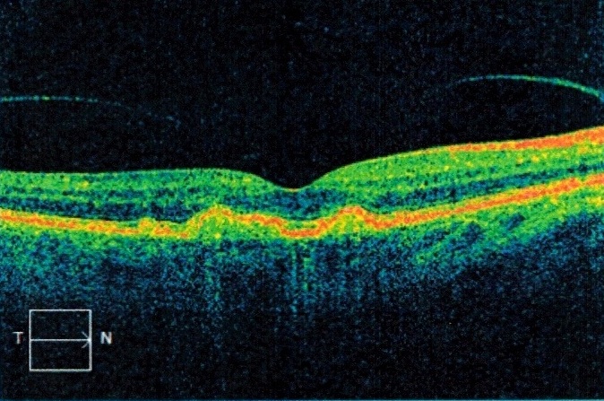
Early dry macular degeneration (AMD) presents itself as bumps in the Retinal Pigmented Epithelium (red layer). Diabetes and bleeding in the eye show up as dark pockets in the inner layers of the macula. This technology is the only way to monitor glaucoma, macular diseases and diabetes to ensure proper treatment appropriate referral if needed. The OCT can detect if Dry AMD is converting to WET AMD before bleeding occurs allowing for prompt referral to a retinal specialist.
HUMPHREY HFA-3 VISUAL FIELDS
The Zeiss Humphrey Visual field testing (perimetry) is still the standard method of following glaucoma and will tell us if the eye is being damaged from glaucoma before it becomes noticeable to the patient. This is the test that presents a series of lights in your peripheral vision and maps out the consistently dimmest light the patient can see at over multiple points in the field of vision, so it can be accurately compared from test to test.
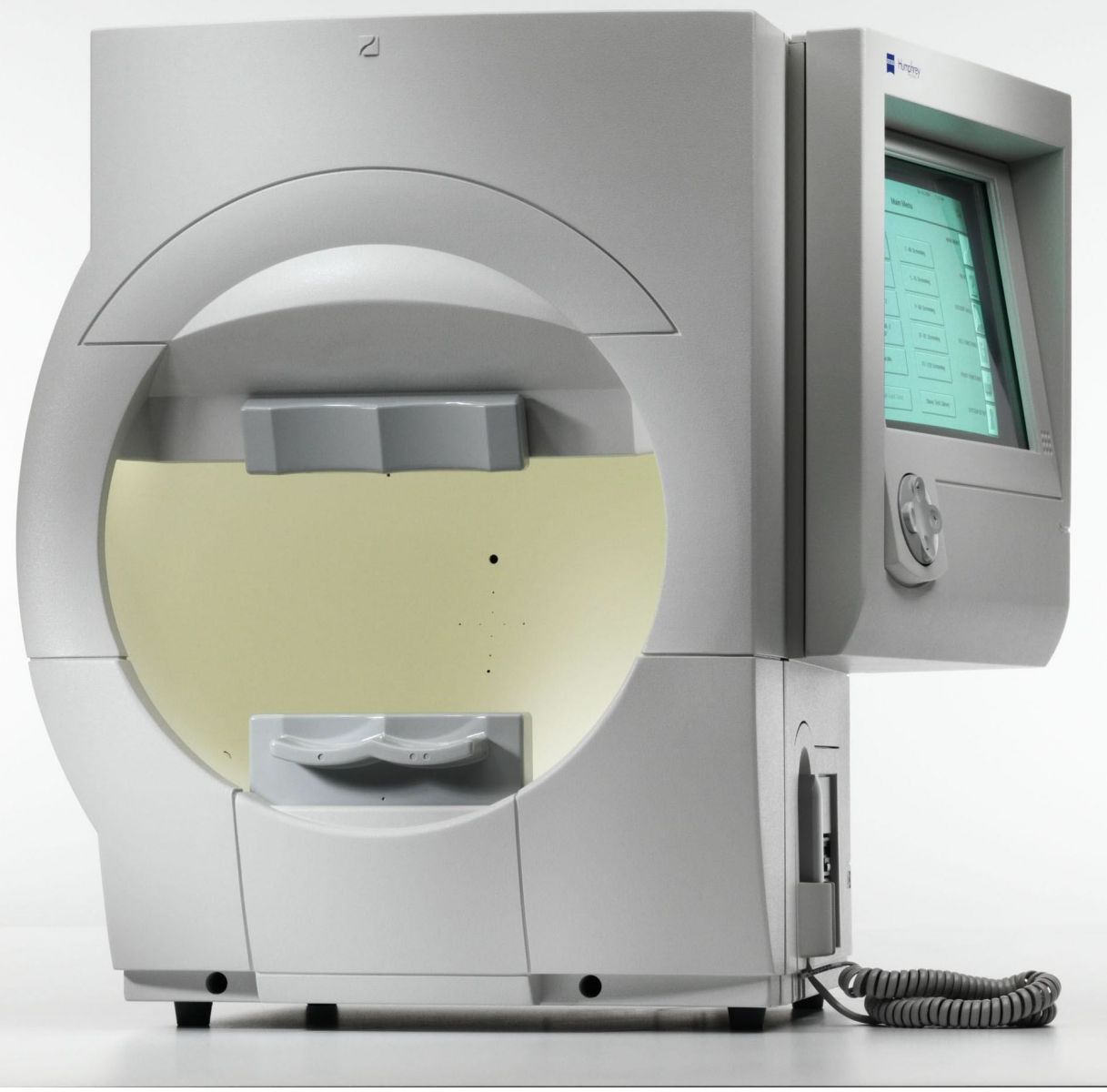
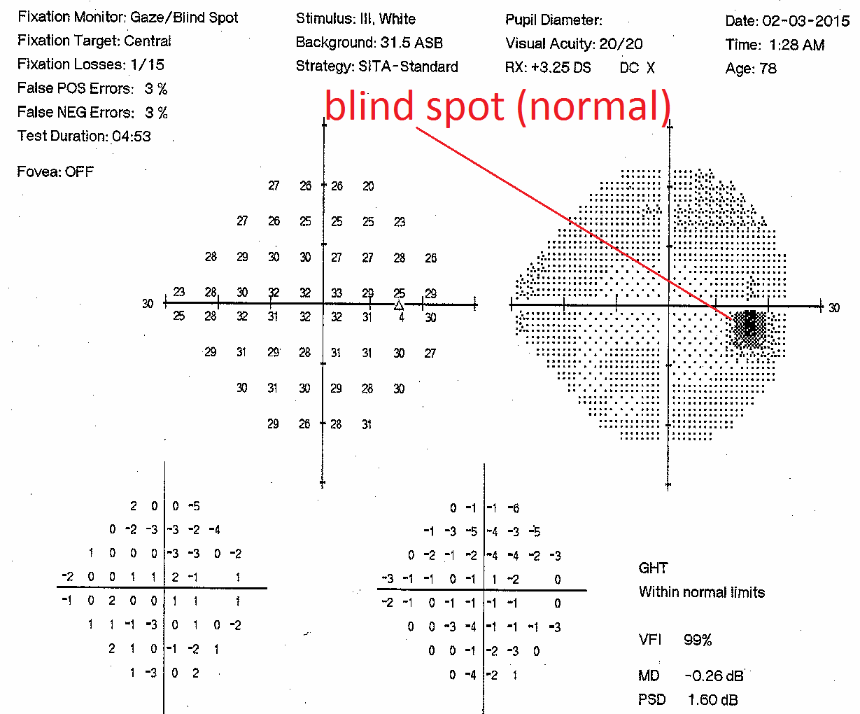
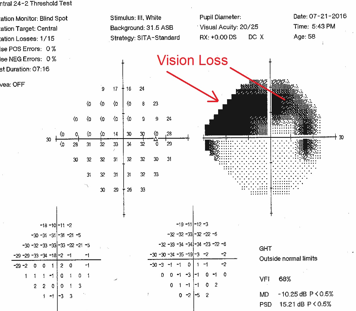
A normal Visual field and one showing permanent vision loss from glaucoma

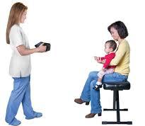
Welsh Allyn portable Auto Refractor is a fun, easy way to obtain prescription results for infants and toddlers. It takes just seconds to determine if your child is nearsighted, farsighted, has an astigmatism, or is at risk for a lazy eye (amblyopia). And they are much more at ease sitting on your lap. It's also a great tool to use on the very elderly who are confined to a wheel chair and may have difficulty answering questions. Please know that our office is 100% handicap accessable, wheelchair friendly.
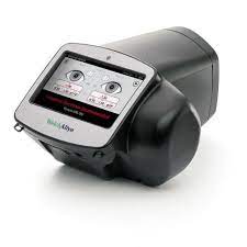
 PORTABLE STILL AND VIDEO EYE IMAGING
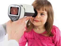 This instrument brings the technology to the patient. It allows for still photography and videography of both the front and the back of the eye. Great for kids and immobile patients. Great for assessing eye infections and styes.
IOL MASTER
 This instrument measures the length of the eye in .01mm increments. We get a baseline reading of all kids who need glasses to help us monitor and control their nearsightedness (myopia). An eye that increases in length becomes nearsighted. Our Myopia Management Program uses a number of techniques to slow down the progression of myopia. Highly nearsighted eyes are at higher risk for Retinal detchment and glaucoma.